Main menu
You are here
Microscopes types
Microscopes types
Various types of microscopes are available for use in the microbiology laboratory. The microscopes have varied applications and modifications that contribute to their usefulness.
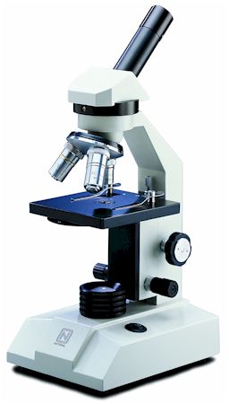
The light microscope. The common light microscope used in the laboratory is called a compound microscope because it contains two types of lenses
that function to magnify an object. The lens closest to the eye is called the ocular, while the lens closest to the object is called the objective. Most microscopes have on their base an apparatus called a condenser, which condenses light rays to a strong beam. A diaphragm located on the condenser controls the amount of light coming through it. Both coarse and fine adjustments are found on the light microscope.
Immersion oil has the same light-bending ability (index of refraction) as the glass slide, so it keeps light in a straight line as it passes through the glass slide to the oil and on to the glass of the objective, the oil-immersion lens. With the increased amount of light entering the objective, the resolution of the object increases, and one can observe objects as small as bacteria. Resolution is important in other types of microscopy as well.
Other light microscopes. In addition to the familiar compound microscope, microbiologists use other types of microscopes for specific purposes. These microscopes permit viewing of objects not otherwise seen with the light microscope.
An alternative microscope is the dark-field microscope, which is used to observe live spirochetes, such as those that cause syphilis. This microscope contains a special condenser that scatters light and causes it to reflect off the specimen at an angle. A light object is seen on a dark background.
A second alternative microscope is the phase-contrast microscope. This microscope also contains special condensers that throw light “out of phase” and cause it to pass through the object at different speeds. Live, unstained organisms are seen clearly with this microscope, and internal cell parts such as mitochondria, lysosomes, and the Golgi body can be seen with this instrument.
The fluorescent microscope uses ultraviolet light as its light source. When ultraviolet light hits an object, it excites the electrons of the object, and they give off light in various shades of color. Since ultraviolet light is used, the resolution of the object increases. A laboratory technique called the fluorescent-antibody technique employs fluorescent dyes and antibodies to help identify unknown bacteria.
Electron microscopy. The energy source used in the electron microscope is a beam of electrons. Since the beam has an exceptionally short wavelength, it strikes most objects in its path and increases the resolution of the microscope significantly. Viruses and some large molecules can be seen with this instrument. The electrons travel in a vacuum to avoid contact with deflecting air molecules, and magnets focus the beam on the object to be viewed. An image is created on a monitor and viewed by the technologist.
The more traditional form of electron microscope is the transmission electron microscope (TEM). To use this instrument, one places ultrathin slices of microorganisms or viruses on a wire grid and then stains them with gold or palladium before viewing. The densely coated parts of the specimen deflect the electron beam, and both dark and light areas show up on the image.
The scanning electron microscope (SEM) is the more contemporary form electron microscope. Although this microscope gives lower magnifications than the TEM, the SEM permits three-dimensional views of microorganisms and other objects. Whole objects are used, and gold or palladium staining is employed.
source: cliffsnotes.com - microscopes types
Copyright© 2010-2012 Grook network
sitemap | Privacy Policy
Follow us on: ![]()
![]()
![]()
![]()
